SAEDNEWS: During ovulation, progesterone increases blood flow and cell growth in the breasts, which is why they may feel slightly firm and sensitive at this time.
According to Saed News, citing Simorgh,The female breast is an important organ, and understanding its anatomy can help identify certain problems and diseases early.
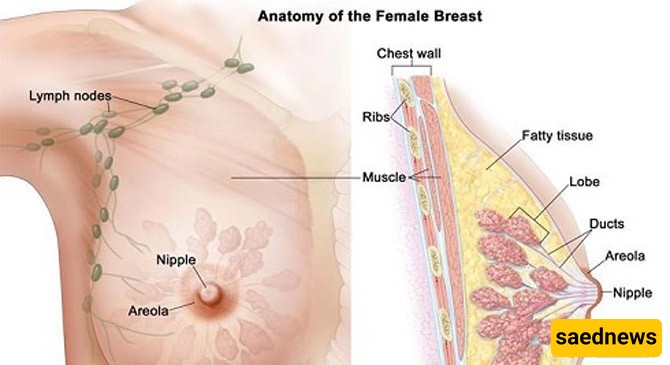
Before puberty, breasts have a darker area called the areola, and in the center of the areola lies a protrusion called the nipple.
Beneath the nipple is a network of fine milk ducts interspersed with fatty tissue. These structures are immature until puberty and are supported by connective tissue called stroma.
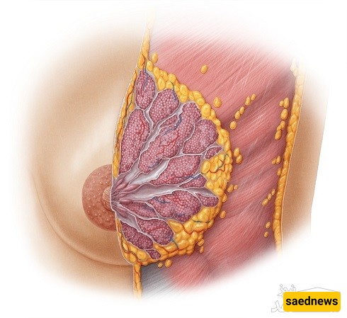
When girls reach puberty, the secretion of female hormones—estrogen and progesterone—increases, prompting the breasts to grow and mature. During this time, the stroma proliferates, milk ducts develop, and an extensive ductal network forms. Inside the breast, lobules begin to develop like buds. Lobules are small sacs that will produce milk after childbirth. Milk from millions of lobules flows through the ducts to the nipple.
Each breast consists of 15–20 sections, arranged like the spokes of a wheel around the nipple. Each section is called a lobe, and each lobe contains smaller units called lobules, which end in small milk-producing glands.
The lobular network connects to the milk ducts, which converge at the nipple to deliver milk during breastfeeding. Most breast cancers originate in the ducts, followed by the lobules and, less commonly, other tissues.

The nipple is centrally located and surrounded by a colored area called the areola. The color of the areola varies among women and can change with hormonal fluctuations, such as menstruation or pregnancy. Nipples may be protruding in some women and slightly inverted in others. During breastfeeding, the areola secretes an oily substance that softens the nipple.
Each breast contains a network of arteries and veins that deliver nutrients and oxygen and remove waste.
In addition to blood vessels, lymphatic vessels are spread throughout the breast, connecting to lymph nodes. Lymph nodes, which feel like small beans upon touch, extend from the breast to the armpits and partially to the collarbone and sternum.
The lymphatic network drains waste, excess fat, proteins, and immune cells toward the heart. It is particularly important in breast cancer because cancer cells can spread through lymphatic pathways. During breast cancer surgery, nearby lymph nodes are often removed and examined. If no cancer cells are found, the cancer is likely in its early stages.
Fat surrounds the lobes of the breast, giving it shape, firmness, and protection from physical impact. While breasts lack muscular tissue, the underlying pectoral muscle helps support them.
Breast function is influenced by estrogen and progesterone, secreted by the ovaries. Estrogen opens milk ducts, while progesterone increases the number of lobules in preparation for breastfeeding. Around ovulation, progesterone increases blood flow and cellular growth in the breasts, which can make them slightly firm and tender.
Breasts are rarely perfectly symmetrical and may fluctuate in size throughout the month. Breast tissue also changes with age. Young women typically have more glandular tissue, making their breasts firmer. After menopause, fatty tissue gradually replaces glandular tissue, reducing firmness. Glandular-dense breasts are harder to evaluate with mammography, which is why mammograms are less commonly used for young women as a screening tool. Older women undergoing hormone replacement therapy may have firmer breasts than expected for their age, which can also complicate mammographic evaluation.