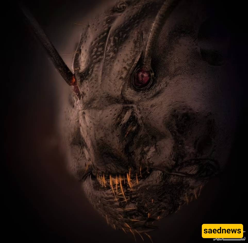
SAEDNEWS: A photographer has captured an unbelievable image of an ant’s face that, at first glance, might make you think it is a scene from a blockbuster horror film. In reality, however, it is a highly detailed and very real close-up photograph of an ant.

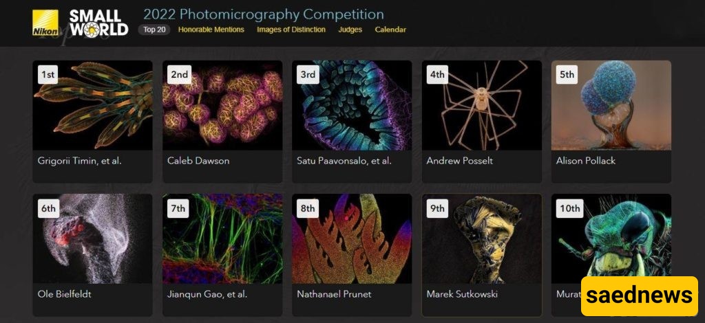
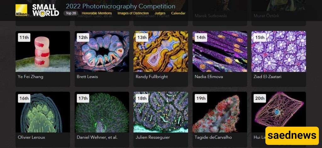
According to Saed News Agency, via Rooziato, a Lithuanian photographer captured an image of an ant under a microscope with 5x magnification. The photo reveals the ant’s red eyes and face in exceptional detail. The image was submitted to Nikon’s Small World Photomicrography Competition and was selected as one of 57 distinguished entries.
Eric Flem, Communications Director at Nikon Instruments, said:
“Every year, Nikon receives a collection of microscopic images that demonstrate remarkable scientific technique and artistry. This year was no exception. At the intersection of art and science, this year’s competition showcased stunning images from scientists, artists, and micrograph photographers from around the world.”
Dr. Kavaliauskas, the photographer of the work, explained:
“I am always looking for details, shadows, and hidden corners. The main goal of photography is to be an explorer. I am fascinated by the masterpieces of creation and the opportunity to see divine patterns.”
While the image of the ant may appear frightening at first glance, Dr. Kavaliauskas emphasized that there is no fear in nature. He said:
“When I first started with microphotography, I thought all insects looked a bit like monsters. But now I am used to it, and I am amazed that so many fascinating, beautiful, and unknown wonders exist beneath our feet.”
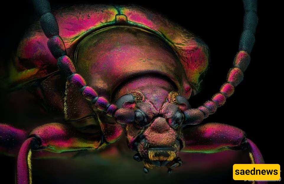
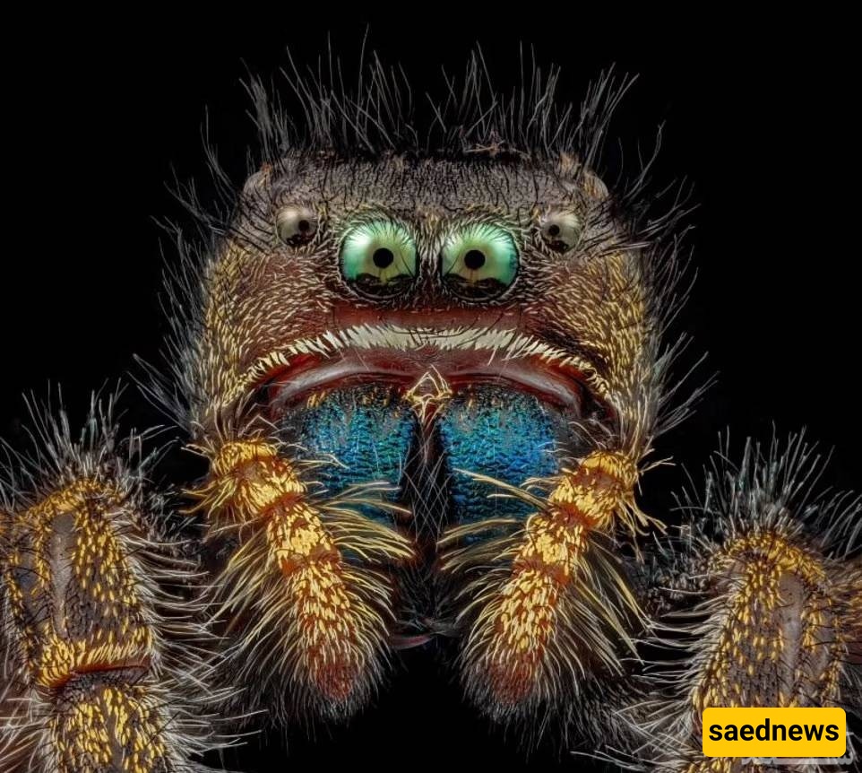
The photographer’s work was not the only close-up insect image featured in the competition. Other selected entries included a striking photo of a red-spotted jewel beetle and a colorful image of a jumping spider.
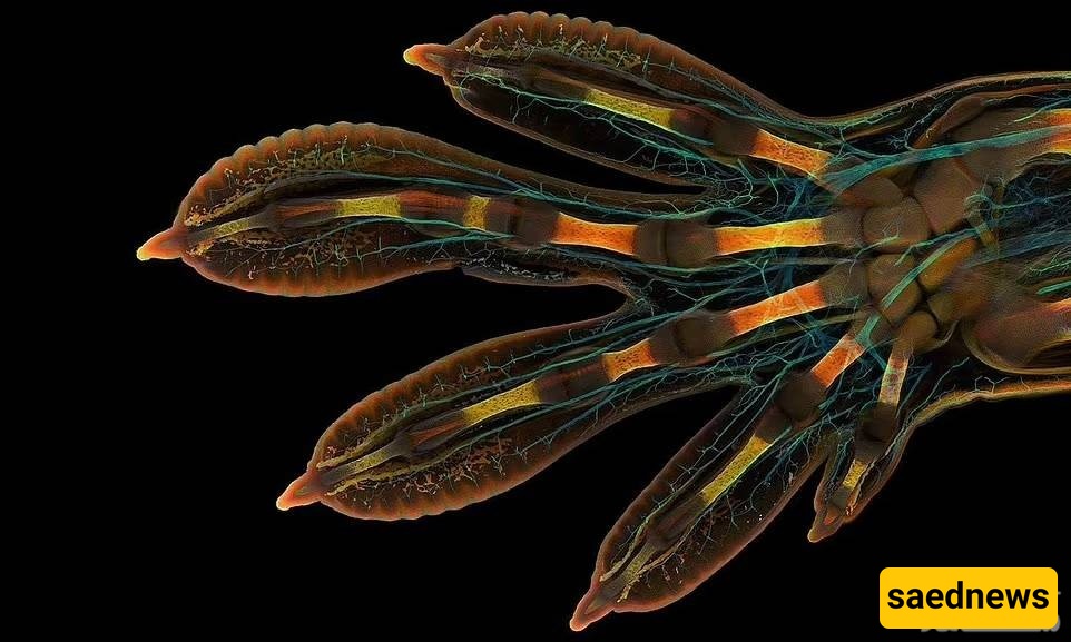
However, the winner of this year’s competition was Gregory Timin, for his remarkable image of a giant gecko embryo hand. Timin said:
“This embryonic hand is about 3 millimeters long, making it a large subject for high-resolution microscopy. The scan consists of 300 sections, each containing around 250 optical slices, requiring more than two days of work and nearly 200 gigabytes of data.”